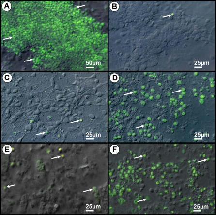
Figure 2. Combined interferential contrast and fluorescent photomicrographs of positive and negative controls (A, B) and Curionopolis- (C, D) or Itacaiunas (E,F)-infected neurons after TUNEL immunolabeling.
Apoptotic nuclei (arrows) are observed in control cultures exposed to UV light (A) and after Curionopolis (D) and Itacaiunas (F) virus inoculation at 4 and 5 days post-inoculation, respectively. Negative control cultures (B) and infected cultures at 1 day post-inoculation (C, E) present few stained nuclei.

