
Figure 2.
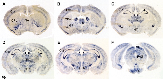
Expression of Cntnap2 mRNA in Postnatal Mouse Brain
Sections of P9 mouse brain were hybridized with a Cntnap2 antisense probe. We detected expression in the cortex (A–D), septum (A), basal ganglia (A and B), many thalamic (B–D) and hypothalamic (C–E) nuclei, with particularly high levels observed in the anterior nucleus and the habenula, part of the amygdala (C), the superior colliculus and the periaqueductal gray (F), pons, cerebellum, and medulla, again with particularly high levels seen in the inferior olive. All panels represent coronal sections and are shown in anterior to posterior order. Ctx, cortex; CPu, caudate putamen; Se, septum; GP, globus pallidus; Th, thalamus; Hip; hippocampal formation; A, amygdala; HTh, hypothalamus; SC, superior colliculus; PAG, periaqueductal gray; Pn, pontine nuclei.