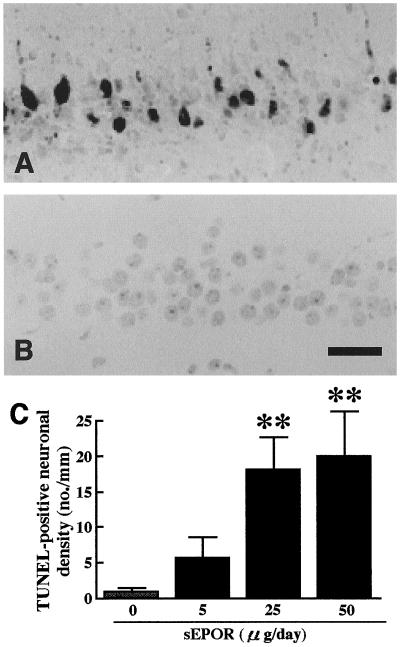
Figure 4.
TUNEL staining of the hippocampal CA1 field of 2.5-min ischemic gerbils after 7 day infusion of sEPOR. (A) An ischemic animal infused with sEPOR at a dose of 25 μg/day; (B) an ischemic animal infused with vehicle. TUNEL-positive neurons were observed only in the ischemic gerbil infused with sEPOR. (C) TUNEL-positive neurons in the hippocampal CA1 field of 2.5-min ischemic animals. The stippled column indicates vehicle-infused ischemic animals and solid columns indicate sEPOR-infused animals. Each value represents mean ± SE (n = 6–8). **P < 0.01, significantly different from the corresponding vehicle-infused ischemic group (statistical significance tested by the two-tailed Mann–Whitney U test). (Bar = 0.1 mm.)

