
Fig. 7. Ultrastructural analyses of mitochondria from B6 and B6.kd/kd mouse kidney and liver.
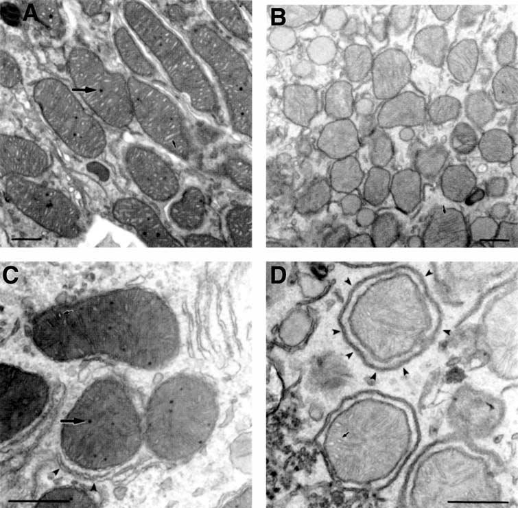
(A Electron micrograph of mitochondria from B6 mouse kidney. Many normal mitochondrial matrix granules are seen (large arrow), as well as well-defined cristae, which are moderately dilated (small arrow). The matrix is homogenous and the overall morphology is smooth and generally oblong. (B) Electron micrograph of mitochondria from B6.kd/kd mouse kidney demonstrating a lack of normal mitochondrial inclusions. In addition, the cristae (small arrow) are compressed, and the matrix is pale and granular. The overall morphology of the B6.kd/kd mitochondria appears smaller and more angular than mitochondria from B6 mice. (C) Electron micrograph for mitochondria from B6 mouse liver demonstrating normal matrix granules (large arrow), and clearly defined, moderately dilated cristae (small arrow). Endoplasmic reticulum (arrow heads) is often closely associated with the mitochondria. The mitochondrial matrix is homogenous and moderately dense. d, Electron micrograph of mitochondria from B6.kd/kd liver demonstrating characteristics similar to those of B6.kd/kd kidney mitochondria. There is an absence of normal matrix granules, and the matrix is paler and more granular than that of B6 mouse liver mitochondria. The cristae (small arrow) seem less affected and appear near normal. The overall mitochondrial morphology again appears generally more angular, but perhaps less so than in B6.kd/kd mouse kidney samples. The endoplasmic reticulum (arrow heads) often appears to completely surround the mitochondria (scale bars, 500 nm).