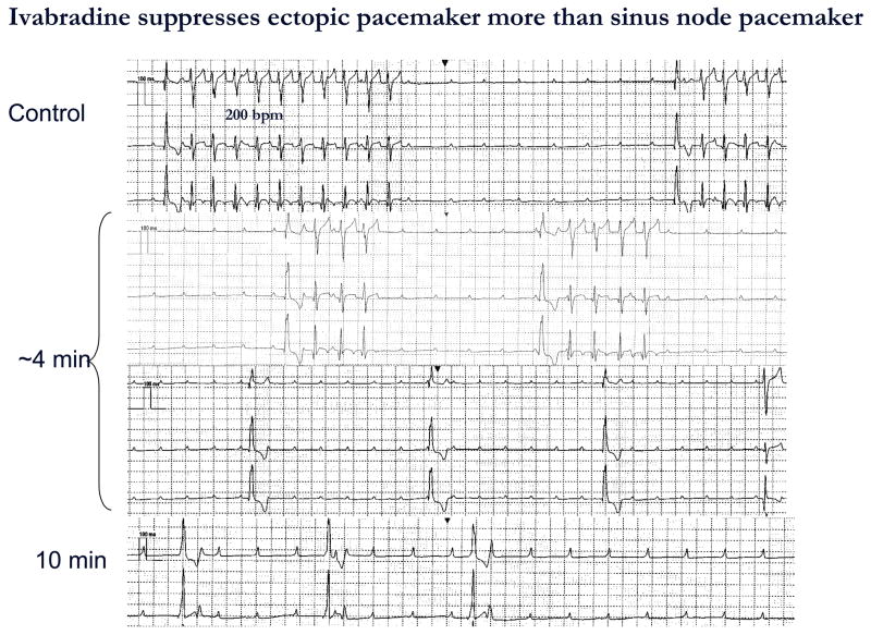
Figure 4.
ECG recorded before, during, and after IVB administration from a dog in complete AV block implanted with HCN212 chimera. Each panel shows continuous traces. The top panel depicts bursts of VT having a rate of 200 bpm and a QRS configuration that pace-mapped to the implantation site. Four min after completing IVB infusion (1 mg/kg IV) over 5 min (middle panel) VT slowed and bursts were shorter. Ten min after IVB infusion (bottom panel) a slow idioventricular rhythm having a wider QRS complex and apparently originating from another site was observed. Atrial rate remained stable during the entire period of observation. ECGs recorded with electronic pacemaker turned off and at paper speed = 25 mm/sec

