
Fig. 1.
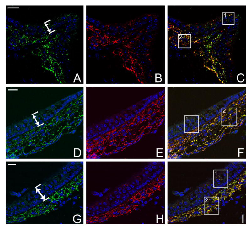
Results of double immunolabeling of Cx26 (red) and Cx30 (green) in semithin sections of vestibular sensory organs of WT adult mice. Cryosections were counterstained with DAPI (blue) to reveal cell nuclei. A,D,G: Immunolabeling of Cx30 in ampulla, saccule, and utricle, respectively. B,E,H: Same sections as in A, D, and G double immunolabeled with Cx26 in ampulla, saccule, and utricle, respectively. C,F,I: Overlapped images of double immunolabeling in the three vestibular sensory regions. Double-headed arrows in A, D, and G indicate the bottom and top borders of the vestibular sensory epithelia. Boxed areas are shown in Figure 2 at higher magnification. Scale bar = ∼20 μm in A (applies to A–C); D (applies to D–F); G (applies to G–I).