
Fig. 2.
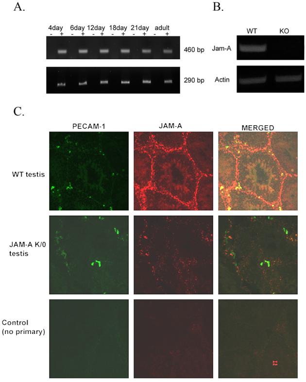
Jam-A transcription and protein localization in WT testis but not in Jam-A null mice. A. RT-PCR results of Jam-A expression in various developmental stages of WT testis. RNA was subjected to first strand synthesis with (+) or without (−) reverse transcriptase, followed by PCR amplification. The expected 460 bp fragment of Jam-A cDNA was detected in every stage (upper panel). Actin is used as control to show the same amount of RT product was used (lower panel). B. Jam-A transcription is detected in WT testis but not in Jam-A null testis. C. Confocal microscopy of JAM-A in mouse testis: Cryostat sections of testis from WT (top panel) and JAM-A null mice (middle panel) were incubated with either pAb JAM-A and mAb against PECAM-1 or no primary antibody for the control (lower panel). Primary antibodies were detected with Alexafluorconjugated secondary antibodies. The bar represents 20 μm.