Figure 1.
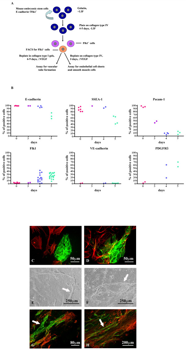
ES cell differentiation into Flk1+ mesodermal, endothelial and smooth muscle cells. A. Schematic showing the protocol for differentiation of CCE cells to mesodermal lineages. B. Expression of cell surface markers at day 0 and days 3–5 of differentiation; cells were analysed by FACS for the markers shown. C-D. Flk1+ cells were plated on collagen type IV in the presence of VEGFA. Cells were double immunostained for VE-cadherin (green) and αSMA (red) (C) or PECAM-1 (green) and αSMA (red) (D). EC sheets were found surrounded by SMCs. E-F. Phase contrast micrographs of Flk1+ cells after FACS sorting and seeding into collagen type I gels in the presence of VEGFA for 6 days. Vascular tube structures are arrowed. G-H. Collagen I gels were sectioned and stained for Pecam-1 (green) and αSMA (red) and showed Pecam-1+ tubular structures (arrows) surrounded by smooth muscle cells.

