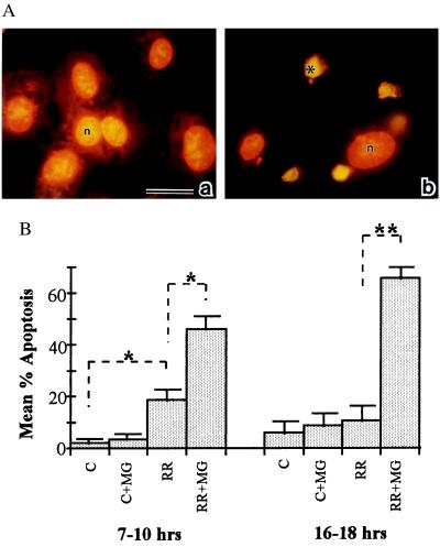
Figure 1.
(A) In situ detection of EC apoptosis during R. rickettsii infection. Infected human umbilical vein ECs were incubated in the presence (b) or absence (a) of the NF-κB inhibitor MG 132. At 18 hr, ECs were fixed and processed for in situ detection of DNA fragmentation by TUNEL and then counterstained with propidium iodide. When visualized under dual wavelength fluorescence, normal nuclei exhibited orange fluorescence (n), whereas apoptotic nuclei displayed green fluorescence and appeared condensed (∗). MG 132 treatment did not induce apoptosis in uninfected ECs (data not shown). (Bar = 20 μm.) (B) The percentages of apoptotic ECs were determined by scoring cells as apoptotic (exhibiting green fluorescence) or normal (exhibiting orange fluorescence) in randomly chosen microscopic fields. Fifteen hundred to 3,000 cells were scored per experimental condition. P values comparing mean percent apoptotic cells between experimental conditions indicated by brackets were determined by Student’s t test and are indicated by asterisks (n = 3 to 5). ∗, P < 0.05; ∗∗, P < 0.00005.

