Figure 2.
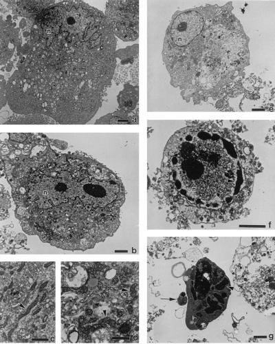
Transmission electron micrographs of R. rickettsii-infected ECs. Shown are uninfected ECs (a and c) or ECs infected for 38 hr in the absence (b and d) or presence (e–g) of MG 132. EC nuclei are denoted by n. Arrowheads in c and d point to mitochondria. Arrowheads in f and g point to areas of condensed chromatin; the arrow in g points to an apoptotic body. [Bars = 2.5 μm (a, b, and e–g) and 1 μm (c and d).]

