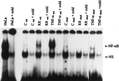
Figure 4.
Gel shift analysis of NF-κB induction in HEF cells. Gel shift analysis was performed on nuclear extracts prepared from control (C), R. rickettsii-infected (RR), and TNF-α-treated (10 ng/ml) wild-type (wt) and mutant (mut) HEFs incubated for 3 hr. Complexes were analyzed on 4% nondenaturing polyacrylamide gels followed by autoradiography. HeLa cells were included as positive markers. Other symbols are as in Fig. 3.

