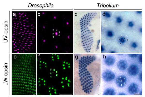
Figure 2.
Differential expression of opsin paralogs in Drosophila and Tribolium. (a and b) Digital sections of Drosophila pupal tissue stained with antibodies against UV-sensitive Rh3 and Rh4 opsins. (a) Low magnification overview. (b) High magnification view. Numbers indicate photoreceptor cell subtypes R1-8. (c and d) Tribolium UV-opsin expression detected by in situ hybridization. (c) Low magnification overview of UV-opsin expression throughout entire pupal retina. (d) High magnification view of cell specific expression of Tribolium UV-opsin. Numbers indicate photoreceptor cell subtypes. (e and f) Digital sections of Drosophila pupal tissue stained with antibody against the long wavelength-specific opsin Rh1. (e) Low magnification overview. (f) High magnification view. Numbers indicate photoreceptor cell subtypes R1-8. (g and h) Tribolium LW-opsin expression detected by in situ hybridization. (c) Overview of LW-opsin expression throughout entire pupal retina. (d) High magnification of cell specific expression of Tribolium LW-opsin. Numbers indicate photoreceptor cell subtypes. Scale bars correspond to 10 μm.

