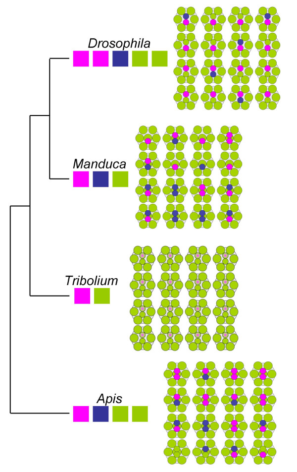
Figure 3.
Comparison of insect photoreceptor mosaics. Schematic presentation of photoreceptor sensitivity arrays in the compound eyes of honeybee (Apis), tobacco hornworm moth (Manduca), Drosophila, and Tribolium. Colored boxes underneath genus names indicate number and wavelength specificities of opsins that are known to be expressed in the main retina. Tree visualizes phylogenetic relationships between the insect orders to which the four species belong [51]. In the retinal mosaics, colors indicate photoreceptor light specificities in either the UV (violet), blue or green wavelength range. Drosophila forms eight photoreceptors per ommatidium. The peripheral photoreceptors R1-6 express a LW-opsin. 70% of the Drosophila ommatidia are of the yellow type, in which the central R8 cell express a the LW-opsin paralog Rh6 and the central R7 cells the UV-opsin Rh4. In the remaining pale-type ommatidia, the central R8 cell express the B-opsin Rh5 and the central R7 cells the UV-opsin Rh3 (Fig. 1) [12]. Honeybee and tobacco hornworm moth develop nine photoreceptors per ommatium due to duplication of the R7 cell fate [52]. However, in both species only the two central R7-like more distally located cells exhibit differential opsin expression ranging from UV to LW sensitive opsins. The peripheral photoreceptor cell homologs R1-6 express LW-opsin as does the proximally located central R8 cell homolog. Five different ommatidia types can be distinguished in the tobacco hornworm moth retina, which differ by number of R7-like cells (1–2) or the combination of B- and UV-opsin expressing R-7 cells [12]. In the honeybee, six different ommatidia types occur which either express G-, UV- or B-opsin in both R7-like cells or in any possible combination [24]. In Tribolium, LW-opsin is expressed in all photoreceptor cells. Co-expression of LW- with UV-opsin in R7 is indicated by gradient from violet to green.

