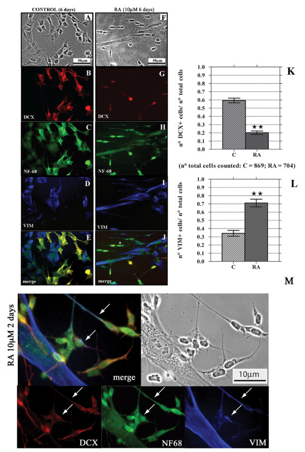
Figure 2.

Six days RA treatment reduces the number of DCX+ cells and increases that of VIM+ cells. Figures here reported are representative of a series of images taken from SK-N-SH cells cultured in control conditions (A-E) or after six days RA treatment (F-J). PC analysis (panels A and F) shows morphological changes induced by RA. After 6 days RA treatment the DCX+ cells are few (G) in comparison to the control (B), and they have the morphology of undifferentiated I-type cells. On the contrary is increased the number of VIM+ cells and the differentiated N-type VIM+ cells do not show immunoreactivity for DCX (I). Bar graphs show the decrease in the number of DCX+ cells (K) and the increase in the number of VIM+ cells (L) after RA treatment. Bars represent mean ± standard error, N = 8, double star = p < 0.01. Neither in control nor in 6 days RA treated samples there are DCX-/VIM- cells immunopositive for both DCX and VIM, but as previously observed in control conditions (see fig.1), some DCX-/VIM- cells are present after RA treatment (see the cell marked with *, panel J). As in the controls (C), NF-68 localizes in all RA treated SK-N-SH cells (H). It is important to note that after the first 2 days of treatment it is possible to observe N-type cells (white arrows) that start to express VIM while DCX is disappearing (M).
