Fig. 2.
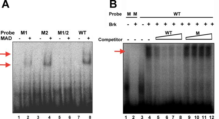
Mad and Brk efficiently and specifically bind the dpp posterior spiracle enhancer. (A) Gelshift with Mad-MH1 domain protein. Lanes 1-8 demonstrate the efficiency of Mad enhancer binding to both sites (shift and supershift indicated with red arrows). Lanes 1, 3, 5, and 7 show the mobility of each oligo alone. Mobility of the WT oligo is significantly impeded in the presence of Mad (Lane 8). Under identical conditions reduced Mad binding to M1 (Lane 2) and M2 (Lane 4) is seen. No binding of Mad to M1+2 is evident. (B) Gelshift with Brk protein. Lanes 1-4 demonstrate the efficiency of Brk enhancer binding (shift indicated with red arrow). Lanes 1 and 3 show the mobility of each oligo alone. The WT oligo is significantly impeded by Brk binding (Lane 4) but the mutant oligo is not (Lane 2). Lanes 4-12 demonstrate the effect of adding increasing amounts of cold competitor oligo (10X, 50X, 100X and 200X) to the WT reaction in Lane 4. Adding cold WT (Lanes 5-8) significantly reduces probe binding at 10X and essentially eliminates binding at 50X. Adding cold M1 (Lanes 9-12) has no effect.

