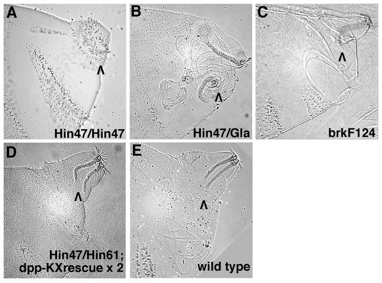
Fig. 4.
Filzkorper defects in dpp and brk mutant embryos suggest that two rounds of Dpp signaling occur during posterior spiracle development. High magnification Nomarski images of embryonic cuticles seen in lateral view. The focus is on abdominal segments 8-11 with the posterior spiracle region of segment 8 indicated by a black arrowhead. (A) dpp null embryos (dppHin47 homozygotes) have an ectopic denticle belt in place of their posterior spiracles. (B) dpp Haploinsufficient embryos (dppHin61 / Gla) have highly disorganized posterior spiracle regions characterized by misshapen Filzkorper that are displaced from their normal location. (C) brkF124 embryos have essentially normal posterior spiracles except that their Filzkorper are U-shaped and do not appear to be connected to the remainder of the tracheal system. (D) dppHin47 / dppHin61 embryos with two copies of the dpp-ΔKX rescue construct also have U-shaped Filzkorper (this view is slightly ventral from true lateral). Wild type embryos have straight Filzkorper that are attached to the spiracular branches (the connection is visible just above the arrowhead).

