
Fig. 1.
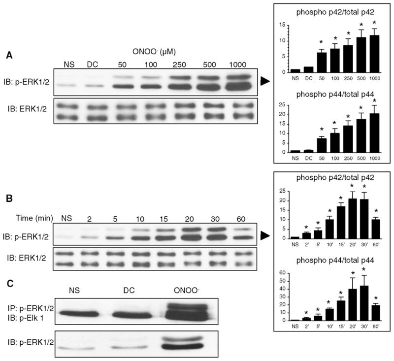
ONOO− induces the phosphorylation and the activity of ERK 1/2 in H9C2 cardiomyocytes. A. Cells were treated with increasing concentrations of ONOO− for 15 min. Phosphorylation of ERK 1/2 was induced at concentrations of ONOO− of 50 μM and above. Decomposed ONOO− (DC) did not phosphorylate ERK. Total ERK was comparable in all the conditions of stimulation. B. Cells were exposed to 500 μM ONOO− for 2–60 min. ERK phosphorylation was maximally induced at 20 min of ONOO− exposure. C. Cells were either not stimulated (NS) or activated with ONOO− (500 μM) or decomposed ONOO− (DC) for 15 min. ERK activity was assessed by Elk-1 phosphorylation after immunoprecipitation of phosphorylated ERK 1/2. As expected, ERK activity correlated with ERK phosphorylation. Densitometric analyses are shown as mean ± S.E.M. of at least four independent experiments. * P < 0.05 vs. NS.