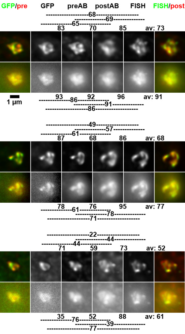
Figure 2.

Comparison of GFP, immunostaining and FISH signals from the same nuclear structure. Upper rows show projections of deconvolved image stacks, lower rows those from non-deconvolved images. As indicated at the top of the panel, color overlays on the left are from GFP (green) and antibody signals before FISH (preAB, red, Cy5), color overlays on the right from antibody signals after FISH (postAB, red, Cy5) and FISH signals (green, FITC). Indicated CC values are based on pair-wise comparisons of 3D image stacks, thus they do not necessarily reflect similarity in the projections. Lines on the side of CC values indicate which signals were compared. Shown are examples for high to average CC values (top and center) and low CC values.
