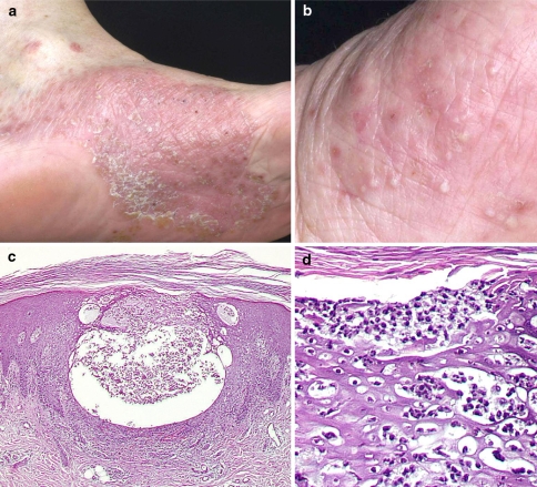
Fig. 1.
Clinical picture of pustulosis palmoplantaris in patient 3 with pustules in different stages of evolution on a sharply delineated erythematous lesion on the left sole (a) and yellowish pustules on the left palm (b). Histological examination showing intraepidermal vesiculopustular dermatitis (c, H.E. stain of a biopsy from the left plantar arch) with intraepidermal accumulation of neutrophils and subcorneal pustule formation (d)

