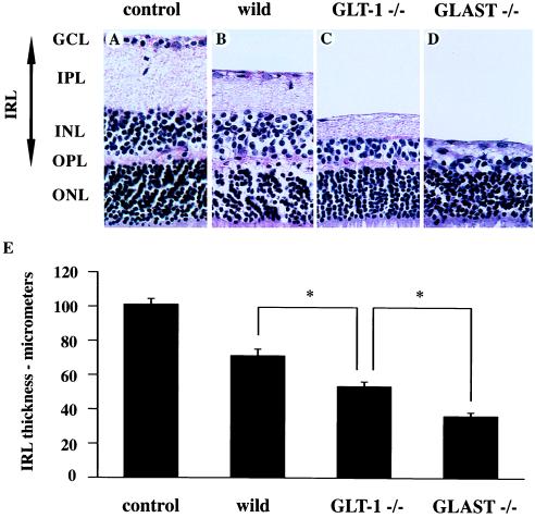
Figure 3.
The ischemic retinal changes in the wild-type and GLAST and GLT-1 mutant mice. (A) Micrograph of a section of a retina in the control eye taken from the wild-type mouse. (B–D) Ischemic retinae from the wild-type (B) and GLT-1 (C) and GLAST (D) mutant mouse. IRL, inner retinal layer; other abbreviations as in Fig. 1. (E) Mean thickness of the inner retinal layers of the retinae in control eyes from the wild-type mice and in ischemic eyes from the wild-type and GLT-1 and GLAST mutant mice. Columns and error bars represent mean ± SEM (∗, P < 0.05).

