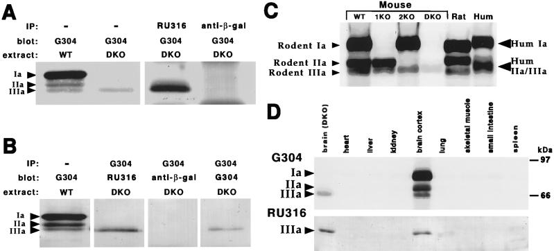
Figure 4.
Distribution of synapsin IIIa protein. Immunoblot analyses of mouse cortex (WT, wild type; 1KO, synapsin I knock-out; 2KO, synapsin II knock-out; DKO, synapsin I/II double knock-out), rat cortex (Rat), and human cortex (Hum) are shown. (A and B) Protein extracts (extract) from WT (50 μg) and DKO (200 μg) brain were loaded in the first two lanes in A or from WT in the first lane B to follow the migration of synapsins Ia, IIa, and IIIa, as indicated by the arrowheads. For immunoprecipitation, the indicated affinity-purified antibody (IP) was incubated with 500 μg of DKO brain protein extract. Antibody–antigen complexes were isolated by using protein A-Sepharose, and the samples were analyzed by immunoblot with the indicated antibody (blot). (C) Expression of synapsins in mouse, rat, and human brain. Protein extracts (50 μg) were loaded in each lane and blots were probed with G304. (D) Expression of synapsins in various mouse tissues. Protein extracts (100 μg) of various tissues were loaded in each lane, and immunoblots were probed with either G304 (Upper) or RU316 (Lower).

