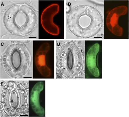
Figure 2.
Localization of ROP2 in V. faba Guard Cells.
(A) An intact V. faba guard cell transformed with RFP-CA-ROP2.
(B) An intact V. faba guard cell transformed with RFP-DN-ROP2.
(C) An intact V. faba guard cell transformed with RFP alone.
(D) An intact V. faba guard cell transformed with GFP-ROP2.
(E) An intact V. faba guard cell transformed with GFP alone.
In all panels, the guard cells at left were transformed by biolistic bombardment and the cells at right were not. A fluorescence image (right) and the corresponding bright-field image (left) of the same cell are shown. The guard cells shown in (D) and (E) were kept under darkness after bombardment until observation, and those in (A) to (C) were irradiated with white light for 3 h before observation. Black dots in bright-field images are gold particles. Focus was on the mid-plane of the cells. Bars = 10 μm.

