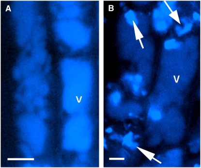
Figure 5.
Localization of UV Fluorescent Material in Wild-Type and Mutant Oat Root Epidermal Cells.
UV confocal microscopy of oat root epidermal cells showing uniform distribution of avenacin A-1 in vacuoles of wild-type roots (A) and patchy distribution of monodeglucosyl avenacin A-1 in sad3 roots (B) (arrow). v, vacuole. Bars = 100 μm.

