
Figure 2. Expression of Crtl1 and versican at ED9.5 and ED10.5.
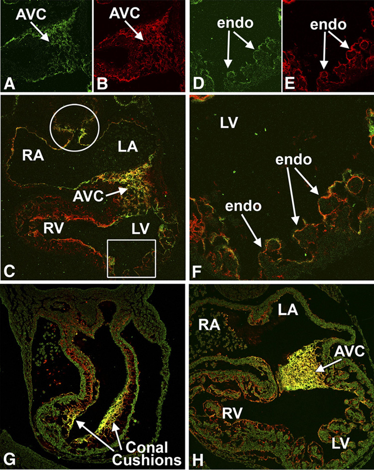
This figure shows immunofluorescently-stained sections co-labeled for Crtl1 (green) and versican (red), (co-exprssion shown in yellow), at ED9.5 (panels A–F) and ED10.5 (G and H). At ED9.5, Crtl1 is expressed in the extracellular matrix surrounding the endocardial and endocardial-derived cells of the AV cushion mesenchyme (panels A and C) and in the lining of the atria (panel C) and ventricles (panels C, D, F). Crtl1 is also expressed in the reflections of the dorsal mesocardium (white circle, panel C). Versican (panels B and E) shows a high degree of co-expression (yellow) with Crtl1 (panels C and F). Panels D–F are higher magnifications of the boxed region in C, showing Crtl1 and versican expression in the extracellular matrix surrounding the endocardial lining of the ventricular trabeculae. At ED10.5, Crtl1 and versican are highly expressed in the sub-endocardial extracellular matrix of the conal cushions in the proximal OFT (panel G). In addition, versican is expressed in the sub-endocardial mesenchyme of the truncal OFT ridges, regions where little Crtl1 expression is detected (panel G). In the AV cushions, Crtl1 and versican also show a high degree of co-expression (panel H). RA=right atrium, LA=left atrium, RV=right ventricle, LV=left ventricle, AVC=atrioventricular cushions, endo=endocardium.