
Figure 8. Reduced levels of versican are implicated in the etiology of the cardiac malformations in the Crtl1-deficient and heterozygous hdf mice.
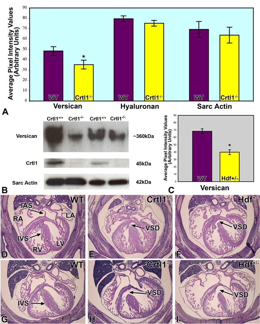
Panel A graphically depicts the levels of versican, hyaluronan, and sarcomeric actin expression, as measured by quantitative immunofluorescence, in the AV cushions of Crtl1−/− mouse embryos at ED13.0. While the levels of versican are significantly reduced (t=2.74; df=8; p=0.012), hyaluronan expression in the AV cushions, and sarcomeric actin expression in the adjacent myocardium, are not significantly changed. Panel B shows Western Blot results, using total hearts of wildtype and Crtl1−/− littermate embryos at ED13.0, confirming the decreased levels of versican in Crtl1−/− mice. Quantification of the western blots indicated a reduction of versican in the Crtl1−/− specimens by approximately 45% (t= 2.66, df= 4, p= 0.02). Crtl1 controls show the absence of Crtl1 in the Crtl1−/− mice, while the sarcomeric actin staining is an independent control of protein loading. Panel C is a graph depicting versican levels in ED13.5 hdf+/− embryos, as measured by quantitative immunofluorescence. The graph demonstrates that versican is significantly reduced, when compared to wild type littermates (p = 0.0005). Panels D–I are hematoxylin-eosin stained sections of wildtype (D and G), Crtl1−/− (E and H), and Hdf+/− (F and I) mice, respectively (D–F: stage ED14.0–14.5; G–I: stage ED15.5–16.0). Panels E, H, F, and I show that Crtl1−/− and Hdf+/− embryos are characterized by very similar ventricular septal defects (black arrows). RA=right atrium, LA=left atrium, RV=right ventricle, LV=left ventricle, IAS=interatrial septum, IVS=interventricular septum, VSD=ventricular septal defect.