
Figure 5.
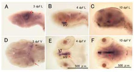
CYP19A2 expression in the brain at 3, 4 and 10 dpf. Embryos were fixed by 4% PFA, and expression of CYP19A2 mRNA in the brain was assessed by whole-mount in situ hybridization. Representative embryos are shown Laterally (A, B, and C) and Ventrally (D, E and F). Arrow indicates CYP19A2 expression in the hypothalamus (HY) and ventral telencephalon (VT), (n=10-12 embryos/age).