
Figure 6.
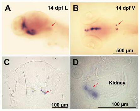
Expression of CYP 19A2 in the adrenal /kidney tissue by whole-mount in situ hybridization. The representative embryos (14 dpf) are shown in Lateral (A) Ventral (B) views and C and D are cross sections of B. D is a higher magnification of C. Arrow indicates expression in the adrenal cells in A, B, C and D (n=10-12 embryos).