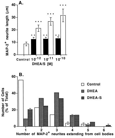
Figure 2.
Dose dependent effect of DHEAS on MAP-2+ neurite outgrowth (A) and effect of steroids on the number of neurites extending from cell bodies (B). (A) Each column shows the mean length of MAP-2+ neurites for at least 100 labeled neurons/condition from three separate wells in one representative experiment. Open bars are DHEAS treatment and solid bars are DHEA treatment. Error bars are ±SD. Neurite length was quantitated by using the nih image 1.57 program. ∗∗∗, P < 0.0001, and ∗∗, P < 0.001 using a one-way ANOVA, with a Scheffe’s post hoc analysis. Three independent experiments gave similar results. (B) Histogram representing the percentage of cells with a specific number (1–6) of MAP-2+ neurites was used to depict data from 10−11 M DHEA and 10−11 M DHEAS treatments. Experiments were performed at 10−12–10−10 M DHEA and DHEAS, and the original data were analyzed by one-way ANOVA, by using Scheffe’s post hoc analysis to determine individual differences at each concentration of steroid. ANOVA showed that DHEA and DHEAS treatments significantly increased the number of MAP-2+ neurites extending from cell bodies compared with untreated control cells. No significant differences were found among the different doses of DHEA and DHEAS, or between DHEA and DHEAS treatments. Open bars are control untreated cells, dark gray bars are DHEA treatment, and hatched bars are DHEAS treatment.

