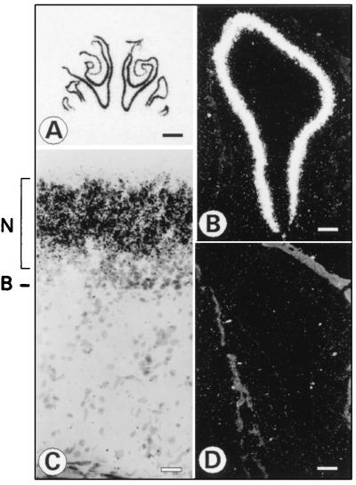
Figure 4.
Expression of CNG4.3 in rat olfactory epithelium. (A) Coronal section through the rat nasal cavity hybridized with a 35S-labeled antisense RNA probe specific for CNG4.3. The nasal septum is in the center with the two nasal cavities on either side. Hybridization is seen throughout the entire extent of the olfactory epithelium. Autoradiographic exposure was for 6 days. The scale bar at bottom right equals 1 mm. (B and D) Darkfield photographs of consecutive sections hybridized to CNG4.3 specific antisense (B) or sense probe (D). Labeling of the olfactory epithelium only is seen with the antisense probe (B). The scalebar in (B and D) equals 140 μm. (C) High magnification brightfield photograph of a slide hybridized to the CNG4.3 specific antisense probe. A uniform signal of black silver grains is observed within the neuronal layer (N) of the olfactory epithelium. B, basal membrane. The scale bar equals 30 μm.

