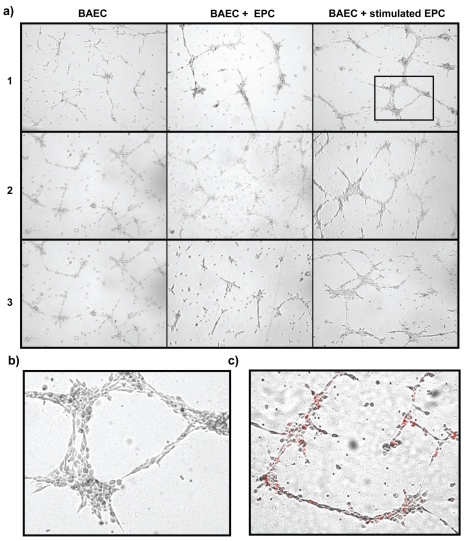
Figure 4.
(a) Coincubation of BAECs with EPCs in an in vitro tube formation assay. Data are shown for EPCs of three healthy volunteers. Whereas unstimulated EPCs only marginally increased tube formation (middle column), EPCs incubated with human insulin in the EPC-CFU assay increased tube formation and led to vascular-like structures after 12 h. (b) Digitally enlarged section of the far right picture in volunteer 1. (c) Tracking of insulin-stimulated EPCs with a lipophil fluorescent cell marker (CM-DiI) showed incorporation of EPCs into the vascular-like structures. A representative picture is shown as an overlay image of light and fluorescence microscopy.

