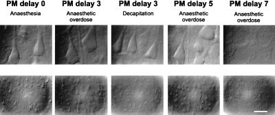
Figure 1.
Layer V pyramidal cells from neocortical slices prepared after various PM delays and visualized with infrared videomicroscopy. Cell morphology was observed with Nomarski optics at high (Upper) and low (Lower) magnifications (calibration bar = 17.5 and 70 μm, respectively). Experimental conditions from left to right: standard slice (PM delay 0); slice prepared after a PM delay of 3 hr from a rat killed by an anesthetic overdose; PM delay of 3 hr, killed by decapitation; PM delay of 5 hr, killed by anesthetic overdose; and PM delay of 7 hr, killed by anesthetic overdose. Note that in the first four conditions large pyramidal cells have a similar appearance. After a PM delay of 7 hr, healthy cells were no longer observed.

