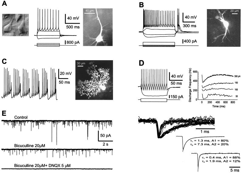
Figure 2.
Cell morphology, intrinsic membrane properties, and synaptic activity in PM rat brain slices. Recordings were obtained from both young ((A–C, E), 16/25-day-old) and adult (D) animals. (A) A neocortical pyramidal cell from layer V was visualized under infrared videomicroscopy (Left), recorded in whole-cell mode (Center; Upper, membrane potential, Lower, injected current) and loaded with biocytin (Right). Resting membrane potential, −71 mV, PM delay of 2 hr. (B) Firing properties (Left) and morphology (Right) of a thalamic relay cell that shows a typical low-threshold calcium spike at the end of a hyperpolarizing current pulse. Resting membrane potential, −71 mV; PM delay of 2 hr. (C) Burst firing behavior induced by steady depolarizing current (Left) in a cerebellar Purkinje cell labeled with biocytin (Right), PM delay of 90 min. (D) Firing properties of an adult neocortical interneuron (fast spiking) from layer II that was spontaneously active. The instantaneous frequency plot shows little accommodation (current pulses of + 50, 100, 150, and 250 pA); PM delay of 2 hr. (E) γ-Aminobutyric acid (GABA) A- and glutamate-mediated synaptic currents recorded in a layer V neocortical interneuron. The cell was voltage clamped at −71 mV and recorded with a pipette containing CsCl. (Left) The spontaneous synaptic currents (Top) were sensitive either to bicuculline (20 μM) or to 6,7-dinitroquinoxaline-2,3-dione (DNQX) (5 μM). (Upper Right) Selected synaptic currents to differentiate slow GABAergic from fast glutamatergic currents. (Lower Right) Both averaged synaptic current types were fitted with two exponentials.

