Figure 3.
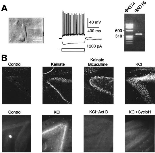
mRNA expression and protein synthesis in PM neurons (PM delay of 1 hr). (A) A vertically oriented interneuron (Left) was recorded in layer V of a neocortical PM slice. This cell fired regularly (Center) and expressed GAD65 mRNAs (Right) as revealed by single-cell RT-PCR. Size markers from bacteriophage φX174 DNA are in the left lane. (B) (Upper) In PM hippocampal slices, synthesis of c-Fos in the dentate gyrus was induced by kainate (10 μM), kainate (10 μM)/bicuculline (20 μM), or by KCl (50 mM) and was revealed by immunocytochemistry. (Lower) Actinomycin D (5 μM) and cycloheximide (35 μM) blocked the induction of c-Fos by high K+.

