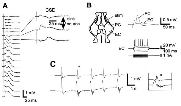
Figure 4.
Cell and network activity in the PM guinea pig. (A) Depth profile analysis in the posterior piriform cortex of an adult guinea pig whole brain in vitro reperfused after a PM delay of 90 min. (Left) Averaged field potentials (n = 5) recorded from surface (top) to interior (60-μm steps) in response to lateral olfactory tract stimulation. (Right) For three field potential traces, the corresponding current source density (CSD) traces are shown. Note the presence of two sinks, the superficial one corresponding to the termination site of the lateral olfactory tract input and the deeper one due to a disynaptic activation of layer II and III pyramidal cells. (B) (Upper) Stimulation of the lateral olfactory tract (stim) evoked synaptic responses in the posterior piriform (PC) and entorhinal (EC) cortices. (Lower) intrinsic membrane properties of a pyramidal cell (intracellular recording) in the EC. (C) In another guinea pig brain (PM delay of 75 min), raising the extracellular K+ concentration to 7.7 mM and adding 200 μM picrotoxin induced bilateral spontaneous and synchronous epileptiform bursts in the temporal lobes.

