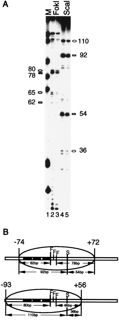
Figure 5.
Translational mapping of the nucleosome on the phas promoter. The AflIII-XbaI fragment (−112 to +141) from the phas promoter in pBluescript II was reconstituted into nucleosome. The resultant nucleosomal complex was treated with micrococcal nuclease (0.07 units), and the kinase-labeled products were loaded onto a 6% native polyacrylamide gel. The relevant band was excised and eluted. The DNA was then extracted with phenol and cleaved with either FokI or ScaI, and the products were separated on a 6% denaturing gel. (A) Two major translational settings were revealed. The FokI (lanes 2 and 3) or ScaI (lanes 4 and 5) fragment positions are denoted by a rectangle (for the −74/+72 setting) or an oval (for the −93/+56 setting). Lane 1 is a marker (M) lane containing MspI-digested pBR322. (B) Diagram of the two major translational positions. Solid rectangles denote TATA boxes. Restriction enzyme sites: F, FokI; S, ScaI.

