Figure 1.
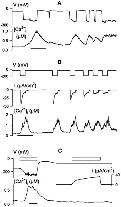
Membrane voltage drives oscillations in cytosolic free [Ca2+] ([Ca2+]i). Concurrent records of voltage, [Ca2+]i, and clamp current (B and C) from three Vicia guard cells bathed in 5 mM Ca2+-Mes, pH 6.1, with 10 mM KCl. Time scales, 1 min. (A) Rise in [Ca2+]i and [Ca2+]i oscillations (lower trace) coincident with spontaneous membrane hyperpolarizations and oscillations in free-running voltage (upper trace). Data from one cell. (B) Cyclic increases in [Ca2+]i (lower trace) evoked under voltage clamp by 20-s steps from −50 to −200 mV (top trace). Clamp current (middle trace) shows inactivation of the inward-rectifying K+ channel current (IK,in) with each [Ca2+]i rise. Note the initial IK,in activation during the first 1–2 s of each step, and the early inactivation of IK,in during the later voltage steps to −200 mV before full [Ca2+]i recovery. (C) [Ca2+]i increase (Left, lower trace) evoked by transfer to 0.1 mM KCl in 5 mM Ca2+-Mes, pH 6.1, to hyperpolarize the free-running voltage (Left, upper trace). No change in [Ca2+]i was observed when membrane voltage was clamped to −75 mV during exposure to 0.1 mM KCl (Right, upper and lower traces). Clamp current (Right, middle trace) was dominated by outward-rectifying K+ channels (not shown; see ref. 20). Open bars (above traces) indicate periods of exposure to 0.1 mM KCl.

