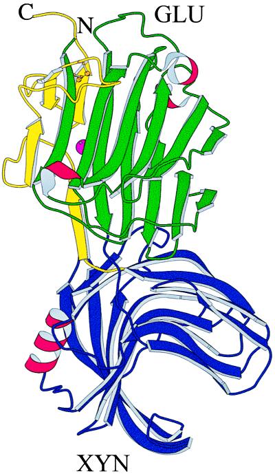
Figure 2.
Schematic drawing of the crystal structure of GluXyn-1. Both polypeptide chain termini are in the GLU domain (Upper) of the insertion fusion protein. In the molscript (40) diagram, the N-terminal part of the GLU domain is colored in green, the XYN domain (Lower) is in blue, and the C-terminal part of the GLU domain, after XYN in the sequence, is in yellow. β-Sheets are shown as curved arrows, α-helices are shown as red, wound ribbons, the calcium ion bound to the GLU domain is shown as a purple sphere half concealed by a β-strand, and the disulfide bridge of the GLU domain is shown in ball-and-stick representation (gold).

