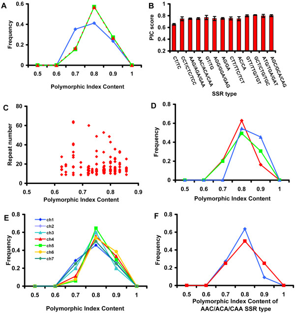
Figure 6.
The polymorphic information content analysis from 129 SSR loci. A. The frequencies of the PIC values of 129 SSR loci are displayed by the SSR type: di-nucleotide SSR (blue diamond), tri-nucleotide SSR (green square), and tetra-nucleotide SSR (red triangle). B. The comparison of PIC scores in the sampled SSRs of different SSR types. The error bars show the standard error of the mean. C. The scatter plot for PIC score and the repeat number among the sampled SSRs. The frequencies of the PIC values of 129 SSR loci are displayed by the genome region (D), exon (red diamond), intron (blue triangle) and intergenic region (green square), and by the chromosome (E). F. PIC score distribution of one SSR type, AAC/ACA/CAA, located either in genic (blue diamond) or intergenic (red square) regions.

