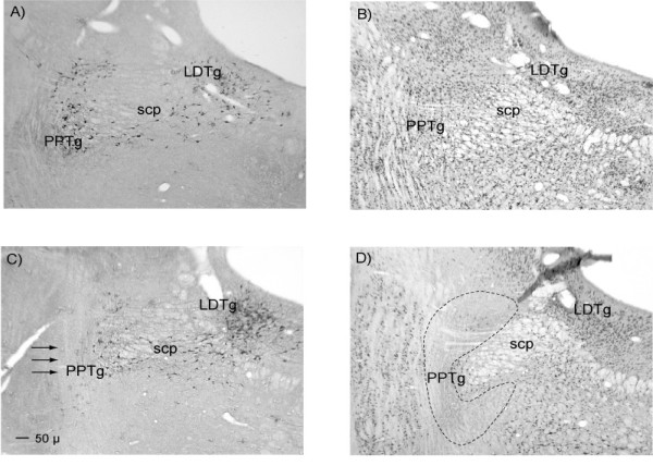
Figure 6.
Photomicrographs illustrating the extent of survival of presumed cholinergic neurons following ibotenate lesions of the PPTg. The four panels show: A – NADPH diaphorase staining in control tissue, B – NeuN/cresyl violet staining in control tissue, C – NADPH diaphorase staining in lesioned tissue with arrows to indicate the expected location of NADPH positive neurons, D – NeuN/cresyl violet staining in lesioned tissue with the borders of the PPTg indicated by black dashed lines. Abbreviations: PPTg = pedunculopontine tegmental nucleus, LDTg = laterodorsal tegmental nucleus, SCP = superior cerebellar peduncle.

