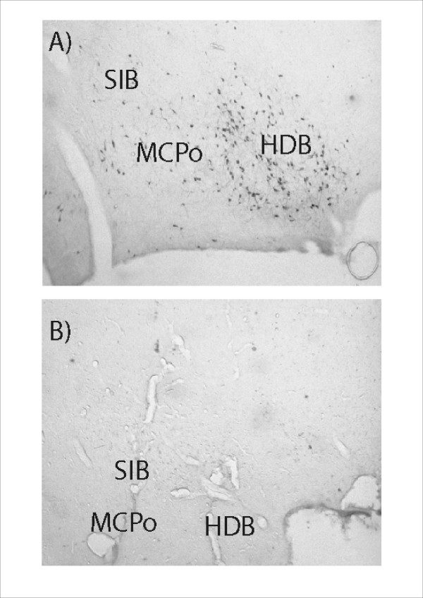
Figure 8.
Photomicrographs illustrating the extent of ChAT positive neuronal loss in the region of the nucleus basalis magnocellularis. The two panels show: A – ChAT positive neurons in a control animal (infusion of Dulbeccos saline), B – Extensive loss of ChAT positive neurons in the same location in an animal who received bilateral infusions of 192 IgG Saporin. Abbreviations: HDB = Horizontal diagonal band of Broca, MCPo = Magnocellular preoptic nucleus, SIB = Substantia innominata basal part.

