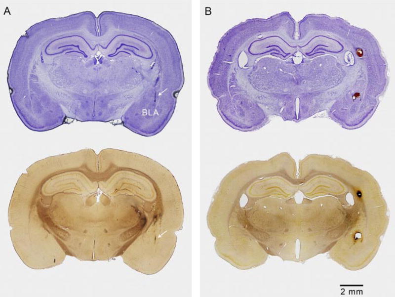
Fig. 6.

CED causes minimal damage in the amygdala and surrounding brain structures in contrast to bolus infusion, which causes cavitation. A, brain obtained from an animal that had been fully kindled and that had received three CED infusions of ω-CTX-G and an additional vehicle infusion. The animal had experienced multiple stimulation-induced kindled seizures. Top, cresyl violet-stained coronal section through the track of the cannula-electrode assembly and right amygdala. Arrow indicates location of track. There is minimal tissue disruption along the track and no damage apparent in the amygdala. Bottom, the adjacent silver-stained section shows some staining in the right striatum, thalamus, and internal capsule, indicating neuronal disintegration. There is no damage apparent in the amygdala. B, brain obtained from a rat infused with 0.05 nmol of ω-CTX-G at a rate of 2.5 μl/min (10-fold greater than for CED). Tracks of the stimulating electrode wires are seen to extend into the right basolateral amygdala and a cavity is present above the nucleus. A second cavity is present in the alveus due to damage from the guide cannula and tracking of the infused solution up the cannula. Cavitation was observed in seven of eight rats treated in a similar manner.
