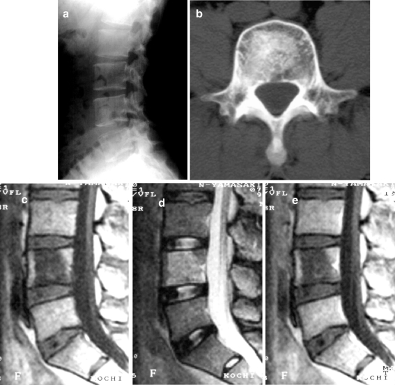
Fig. 1.
A 30-year-old man who complained of low back pain (patient 1). a Lateral radiograph of the lumbar spine reveals vague sclerosis in the fourth lumbar vertebra. b Axial computed tomography (CT) scan of the fourth lumbar vertebra demonstrates significant sclerosis in the center of the body, partly extending to the cortex. c Sagittal T1-weighted spin echo magnetic resonance (MR) image reveals a large intraosseous lesion with low signal intensity. The normal bone marrow signal is preserved in the anterior and posterior portions of the body. d Sagittal T2-weighted MR image shows slightly bright signal intensity intermingled with intermediate signal. e Sagittal gadolinium-DTPA-enhanced MR image does not demonstrate any enhancement. No soft tissue mass is recognized

