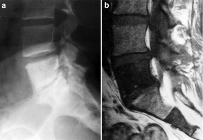
Fig. 3.
A 43-year-old woman who was found to have two separate abnormalities in the lower spine during an imaging study for a traffic accident (patient 7). a Lateral radiograph of the lumbar spine and sacrum reveals intense sclerosis of the entire fifth lumbar vertebral body and mild sclerosis in the cephalad portion of the sacrum. b Sagittal T1-weighted MR image reveals homogeneous low signal intensity in both the fifth lumbar and second sacral vertebrae

