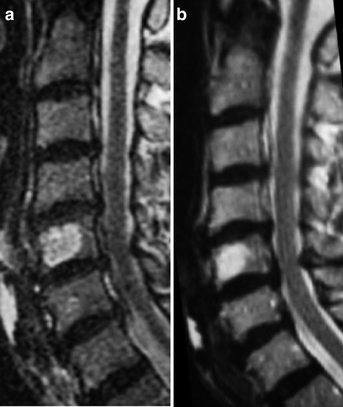
Fig. 4.
A 52-year-old man who complained of mild upper back pain (patient 5). a Sagittal T2-weighted MR image reveals an intraosseous lesion with high signal intensity in the fifth cervical vertebra at initial presentation. b Sagittal T2-weighted MR image demonstrates no progressive disease 14 months after needle biopsy. No extraosseous tumor extension or enlargement is recognized

