
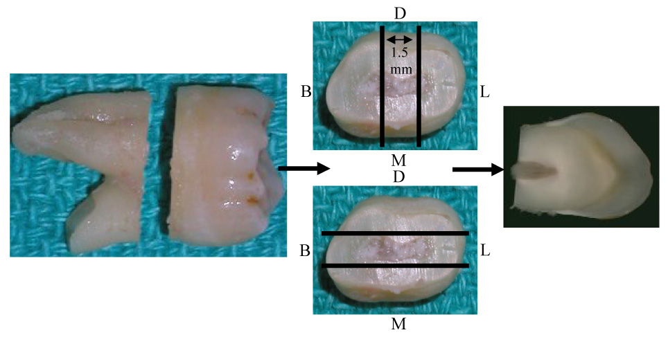
Figure 1.
Sectioning technique for obtaining a 1.5 mm mesio-distal (M,D) or bucco-lingual (B,L)section of coronal tooth structure. Parallel lines indicate approximate area of section. Arrows indicate flow of sectioning procedures.
Official websites use .gov
A
.gov website belongs to an official
government organization in the United States.
Secure .gov websites use HTTPS
A lock (
) or https:// means you've safely
connected to the .gov website. Share sensitive
information only on official, secure websites.
Sectioning technique for obtaining a 1.5 mm mesio-distal (M,D) or bucco-lingual (B,L)section of coronal tooth structure. Parallel lines indicate approximate area of section. Arrows indicate flow of sectioning procedures.