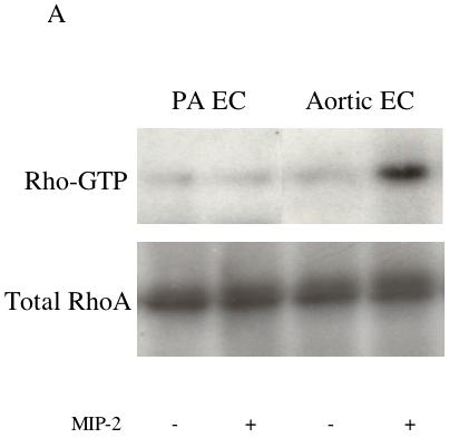
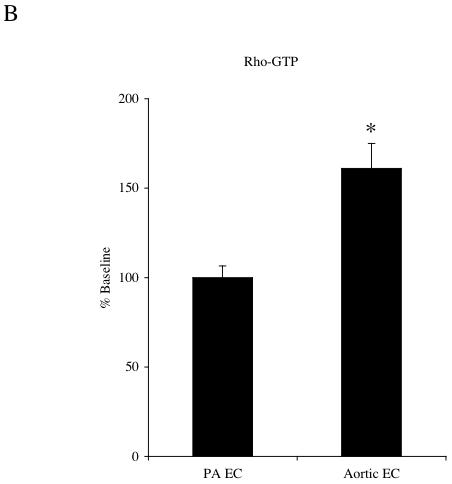
Figure 2.


A: Representative immunoblots of activated Rho-GTP and total RhoA protein in pulmonary artery endothelial cells (PA EC) and aortic endothelial cells (EC) without (-) and with (+) MIP-2 (10 ng/ml) after a 30 min treatment. B: Average increase in RhoA-GTP after MIP-2 relative to baseline control. MIP-2 caused a significant increase in Rho-GTP only in aortic EC (* indicates p = 0.008). Rho-GTP in PA EC was unaffected by MIP-2 treatment (n=5 experiments / cell type).
