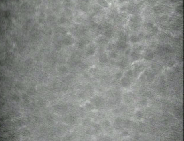
FIGURE 6.

Anterior keratocytes in late endothelial failure (LEF). Confocal microscopy image of anterior keratocytes in a penetrating graft with LEF shows visible cell processes, suggesting activation of keratocytes and preventing identification of individual cells for quantitative analysis in some penetrating grafts. In normal corneas, keratocyte nuclei, and not cell processes, are visible by confocal microscopy.
