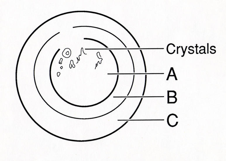
FIGURE 1.
Corneal diagram of location of corneal changes in Schnyder crystalline corneal dystrophy. Initial changes are noted in central cornea (A) with occurrence of corneal crystals and/or central haze followed by formation of arcus lipoides (C) and finally midperipheral stromal haze (B). Reprinted from Weiss JS, et al.34

