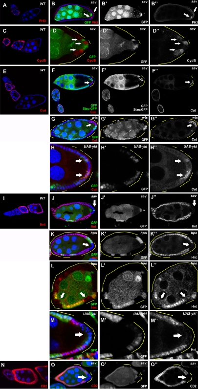
Figure 3. Notch signaling is disrupted in PFC clones of Hpo pathway mutants.
(A and C) PH3 and Cyclin B are expressed sporadically in immature follicle cells during early stages (S1–S6) in wild-type egg chambers. In sav mutants, staining of PH3 (B) and Cyclin B (D) was occasionally found in mutant PFC after stage 6 (arrows). (E) In wild-type egg chambers, Cut is expressed in follicle cells until about stage 6. (F and G) Prolonged Cut expression was found in sav (F) and wts (G) PFC clones at stages 8–10 of oogenesis. (I) Hnt is expressed in follicle cells after stage 6 in the wild type. No Hnt expression was found in sav (J) or hpo (K) mutant PFC in stage-8 egg chambers. (L) Lack of Hnt staining was also occasionally observed in anterior and lateral hpo clones in stage 7 egg chambers (arrows). Overexpression of Yki caused prolonged Cut expression (H) and decreased Hnt expression (M). (N) The E(spl):CD2 Notch activity reporter, visualized by CD2 staining, is upregulated in follicle cells during stages 7–10A of oogenesis in wild-type egg chambers. (O) Lack of CD2 staining was observed in sav mutant PFC in this stage-7 egg chamber (arrow).

