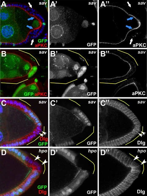
Figure 7. Apical-basal polarity in Hpo-defective follicle cells.
sav mutant follicle cells that remain in contact with the germline display correct localization of the apical marker, aPKC (A, blue arrows; compare to B which shows wildtype cell pattern (GFP-positive cells) as well as no obvious defects in neighboring clone cells). sav clone cells in outer layers of a multilayered clone, however, do not show apical accumulations (A, white arrows). Localization of the basal-lateral marker Dlg appears largely correct in both sav (C) and hpo (D) clones (white arrowheads), but see Results for further description.

